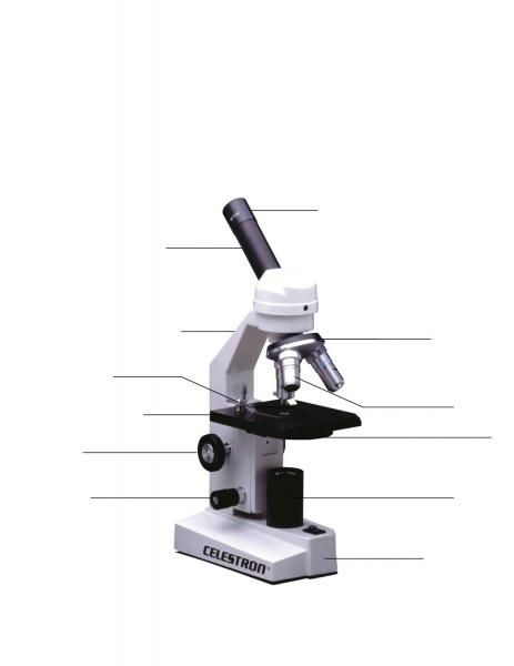
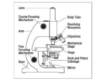
44 microscope picture with labels
What is Electron Microscopy? - UMASS Medical School Conventional scanning electron microscopy depends on the emission of secondary electrons from the surface of a specimen. Because of its great depth of focus, a scanning electron microscope is the EM analog of a stereo light microscope. It provides detailed images of the surfaces of cells and whole organisms that are not possible by TEM. Microscope Types (with labeled diagrams) and Functions The working principle of a simple microscope is that when a lens is held close to the eye, a virtual, magnified and erect image of a specimen is formed at the least possible distance from which a human eye can discern objects clearly. Simple microscope labeled diagram Simple microscope functions It is used in industrial applications like:
Parts of a Microscope Labeling Activity - Storyboard That Create a poster that labels the parts of a microscope and includes descriptions of what each part does. Click "Start Assignment". Use a landscape poster layout (large or small). Search for a diagram of a microscope. Using arrows and textables label each part of the microscope and describe its function. Copy This Storyboard* More options
Microscope picture with labels
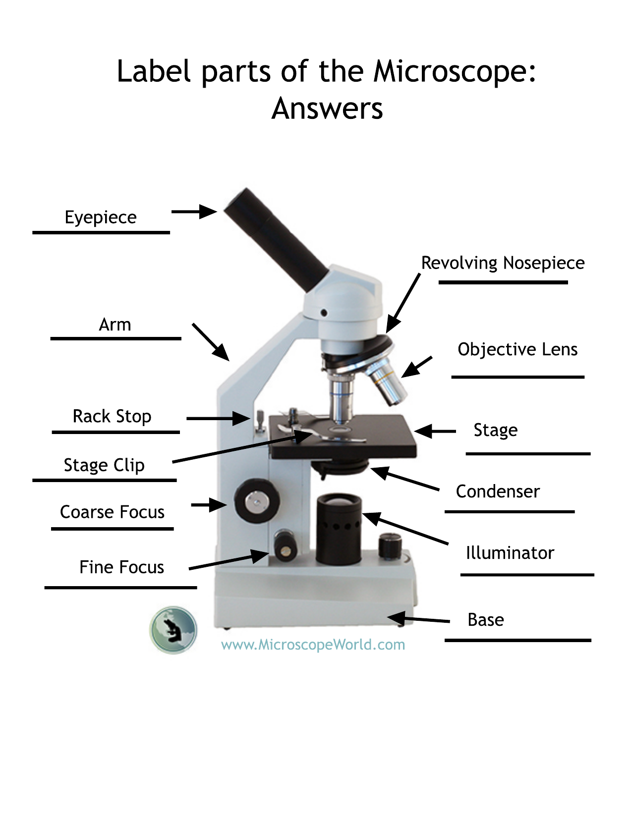
Looking at the Structure of Cells in the Microscope ... The phase-contrast microscope and, in a more complex way, the differential-interference-contrast microscope, exploit the interference effects produced when these two sets of waves recombine, thereby creating an image of the cell's structure . Both types of light microscopy are widely used to visualize living cells. PDF Label parts of the Microscope Label parts of the Microscope: . Created Date: 20150715115425Z LSM 900 with Airyscan 2 – Compact Confocal Microscope for ... In microscopy, this translates into the best contrast and resolution while maintaining minimum light exposure. LSM 900, your compact confocal microscope, provides this with components optimized to deliver the best imaging results. Get high-end confocal imaging in a small footprint. Improve any confocal experiment with LSM Plus.
Microscope picture with labels. Home: The Histology Guide You can see histological slides on the pages and can turn labels on or off to help them identify features. In some cases, there is a section like a 'virtual microscope' - you can scan around a large picture using the mouse and try to identify features. This emulates as closely as possible the experience of using a microscope. Looking at the Structure of Cells in the Microscope A typical animal cell is 10–20 μm in diameter, which is about one-fifth the size of the smallest particle visible to the naked eye. It was not until good light microscopes became available in the early part of the nineteenth century that all plant and animal tissues were discovered to be aggregates of individual cells. This discovery, proposed as the cell doctrine by Schleiden and … ZEISS Axioscan 7 Microscope Slide Scanner Digitize your specimens with Axioscan 7 – the reliable, reproducible way to create high-quality virtual microscope slides. Axioscan 7 combines qualities that you would not expect to get in a slide scanner: high speed digitization and outstanding image quality plus an unrivaled variety of imaging modes are all available in a fully automated and easy to operate system. What is Electron Microscopy? - UMASS Medical School Because the size of the raster at the specimen is much smaller than the viewing screen of the CRT, the final picture is a magnified image of the specimen. Appropriately equipped SEMs (with secondary, backscatter and X-ray detectors) can be used to study the topography and atomic composition of specimens, and also, for example, the surface distribution of immuno-labels.
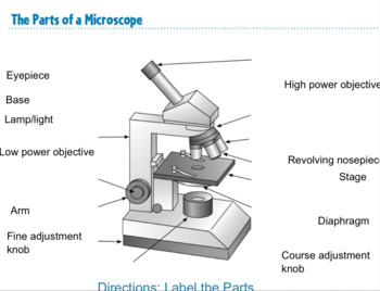
Parts of a Simple Microscope - Labeled (with diagrams) A simple microscope is a very first type of microscope ever created. It consists of simple parts and performs simple functions. Although there are now many advanced microscope types, some applications may still demand the use of a simple microscope. In this article, we are going to discuss the parts and functions of a simple microscope. Microscope Label Worksheets & Teaching Resources | TpT Label the Microscope by Crista Tiboldo 19 FREE PDF (178.64 KB) Worksheet identifying the parts of the compound light microscope. Answer key: 1. Body tube 2. Revolving nosepiece 3. Low power objective 4. Medium power objective 5. High power objective 6. Stage clips 7. Diaphragm 8. Light source 9. Eyepiece 10. Arm 11. Stage 12. Home: The Histology Guide You can see histological slides on the pages and can turn labels on or off to help them identify features. In some cases, there is a section like a 'virtual microscope' - you can scan around a large picture using the mouse and try to identify features. This emulates as closely as possible the experience of using a microscope. Micro Module 1 Flashcards | Quizlet Study with Quizlet and memorize flashcards containing terms like Move the terms into the correct empty boxes to complete the concept map., Drag the images and/or statements to their corresponding class to test your understanding of the main types of microbes., Drag the images or descriptions to their corresponding class to test your understanding of the cellular organization …
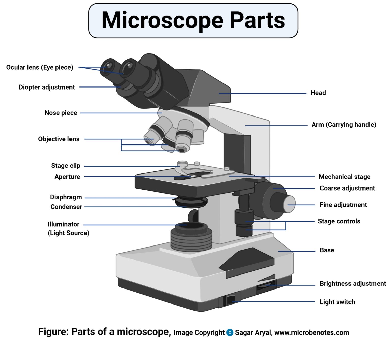
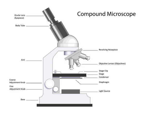
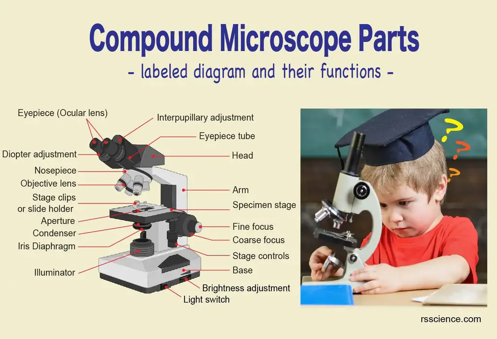
Microscope, Microscope Parts, Labeled Diagram, and Functions Revolving Nosepiece or Turret: Turret is the part of the microscope that holds two or multiple objective lenses and helps to rotate objective lenses and also helps to easily change power. Objective Lenses: Three are 3 or 4 objective lenses on a microscope. The objective lenses almost always consist of 4x, 10x, 40x and 100x powers. The most common eyepiece lens is 10x and when it coupled with ... Simple Microscope - Diagram (Parts labelled), Principle, Formula and Uses The working principle of a simple microscope is that when a lens is held close to the eye, a virtual, magnified and erect image of a specimen is formed at the least possible distance from which a human eye can discern objects clearly. Magnification formula The magnification power of a simple microscope is expressed as: M = 1 + D/F Where Compound Microscope Parts - Labeled Diagram and their Functions The eyepiece (or ocular lens) is the lens part at the top of a microscope that the viewer looks through. The standard eyepiece has a magnification of 10x. You may exchange with an optional eyepiece ranging from 5x - 30x. [In this figure] The structure inside an eyepiece. The current design of the eyepiece is no longer a single convex lens. Cell Size and Scale - University of Utah Smaller cells are easily visible under a light microscope. It's even possible to make out structures within the cell, such as the nucleus, mitochondria and chloroplasts. Light microscopes use a system of lenses to magnify an image. The power of a light microscope is limited by the wavelength of visible light, which is about 500 nm.
Microscope Labeling Game - PurposeGames.com About this Quiz. This is an online quiz called Microscope Labeling Game. There is a printable worksheet available for download here so you can take the quiz with pen and paper. This quiz has tags. Click on the tags below to find other quizzes on the same subject. Science.
Fisherbrand Square Microscope Slide Labels Label size: 0.875 x 0.875 in ... Description. Intended to be used to label microscope slides. Designed to be individually fed into a laser or ink jet printer. Individual labels measure 7/8 x 7/8 in. 40 labels to a sheet, sold in packs of 1,000 labels (25 sheets per pack) This product (s) resides on a Fisher Scientific GSA or VA contract.
Labeling the Parts of the Microscope | Microscope activity, Science ... Description Worksheet identifying the parts of the compound light microscope. Answer key: 1. Body tube 2. Revolving nosepiece 3. Low power objective 4. Medium power objective 5. High power objective 6. Stage clips 7. Diaphragm 8. Light source 9. Eyepiece 10. Arm 11. Stage 12. Coarse adjustment knob 13. Fine adjustment knob 14. Base S
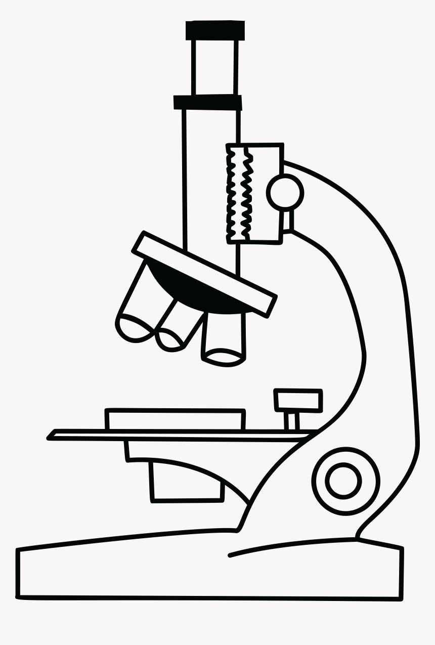
Microscope With Labels clip art | Microscope parts, Scientific method ... Microscope With Labels clip art | Microscope parts, Scientific method, Science diagrams From clker.com vector clip art online, royalty free & public domain Download Clker's Microscope With Labels clip art and related images now. Multiple sizes and related images are all free on Clker.com. D Dixie Tsutsaeva 2k followers More information
Classroom Objects – ESL Flashcards Description. Here are some flashcards for teaching the names of common school supplies and objects usually found in classrooms. The set includes both items that students (should) have in their own school bags as well as items likely to be found …
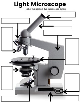
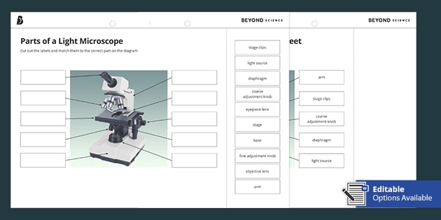
Microscope Labeling - The Biology Corner Students label the parts of the microscope in this photo of a basic laboratory light microscope. Can be used for practice or as a quiz. ... 20. A microscope has an ocular objective of 10x and a high power objective of 50x, what is the microscope's total magnification? _____
Classroom Objects – ESL Flashcards Description. Here are some flashcards for teaching the names of common school supplies and objects usually found in classrooms. The set includes both items that students (should) have in their own school bags as well as items likely to be found on the teacher’s desk only, such as a stapler.
LSM 900 with Airyscan 2 – Compact Confocal Microscope for In microscopy, this translates into the best contrast and resolution while maintaining minimum light exposure. LSM 900, your compact confocal microscope, provides this with components optimized to deliver the best imaging results. Get high-end confocal imaging in a small footprint. Improve any confocal experiment with LSM Plus.
A Study of the Microscope and its Functions With a Labeled Diagram ... To better understand the structure and function of a microscope, we need to take a look at the labeled microscope diagrams of the compound and electron microscope. These diagrams clearly explain the functioning of the microscopes along with their respective parts. ... The camera present within the microscope captures images to reveal the finer ...
Microscope Drawing: How to Sketch Microscope Slides Outline the general shapes: Draw the outline of largest shape onto the paper, making it fit within the quarters. Keep you pencil drawings light and adjust the shape as needed. This may require going between the microscope slide and the drawing in order to get the proportions and shape correct. Now move to the other shapes in your field of view.
The Parts of a Microscope (Labeled) Printable - TeacherVision The Parts of a Microscope (Labeled) Printable. Download. Add to Favorites. Share. This diagram labels and explains the function of each part of a microscope. Use this printable as a handout or transparency to help prepare students for working with laboratory equipment.
Microscope For Sale in Clondalkin, Dublin from Lndnll Microscope, Used Crazy Random ... Personalised ties with any picture and any text you like. €15.00 ... Custom Printed Stickers / Decals / Wedding Labels. €25.00
Microscope Labeled Pictures, Images and Stock Photos Browse 49 microscope labeled stock photos and images available, or start a new search to explore more stock photos and images. Newest results Fluorescent Imaging immunofluorescence of cancer cells growing... Microscope diagram vector illustration. Labeled zoom instrument... Microscope diagram vector illustration.
Label the microscope — Science Learning Hub All microscopes share features in common. In this interactive, you can label the different parts of a microscope. Use this with the Microscope parts activity to help students identify and label the main parts of a microscope and then describe their functions. Drag and drop the text labels onto the microscope diagram.
ZEISS Axioscan 7 Microscope Slide Scanner Digitize your specimens with Axioscan 7 – the reliable, reproducible way to create high-quality virtual microscope slides. Axioscan 7 combines qualities that you would not expect to get in a slide scanner: high speed digitization and outstanding image quality plus an unrivaled variety of imaging modes are all available in a fully automated and easy to operate system.
Microscope For Sale in Clondalkin, Dublin from Lndnll - Adverts.ie Microscope, Used Crazy Random Stuff For Sale in Clondalkin, ... Personalised ties with any picture and any text you like. €15.00 . Personalised Rectangular Metal Photo Keyring. €10.00 ... Custom Printed Stickers / Decals / Wedding Labels. €25.00
Label Microscope Diagram - EnchantedLearning.com Using the terms listed below, label the microscope diagram. arm - this attaches the eyepiece and body tube to the base. base - this supports the microscope. body tube - the tube that supports the eyepiece. coarse focus adjustment - a knob that makes large adjustments to the focus. diaphragm - an adjustable opening under the stage, allowing ...
Microscope Parts, Function, & Labeled Diagram - slidingmotion Microscope parts labeled diagram gives us all the information about its parts and their position in the microscope. Microscope Parts Labeled Diagram The principle of the Microscope gives you an exact reason to use it. It works on the 3 principles. Magnification Resolving Power Numerical Aperture. Parts of Microscope Head Base Arm Eyepiece Lens
Anatomy Chart - How to Make Medical Drawings and Illustrations … Anatomy Chart What is an Anatomy Chart? An anatomy chart refers to a visual depiction of the human body. It can show the entire body or focus on a particular system using systemic anatomy such as the muscular, skeletal, circulatory, digestive, endocrine, nervous, respiratory, urinary, reproductive, and other systems. There are many different branches of anatomy …
Microscope Parts and Functions First, the purpose of a microscope is to magnify a small object or to magnify the fine details of a larger object in order to examine minute specimens that cannot be seen by the naked eye. Here are the important compound microscope parts... Eyepiece: The lens the viewer looks through to see the specimen.
Electron Microscopy Images - Dartmouth Transmission electron microscope image of a thin section cut through the bronchiolar epithelium of the lung (mouse), which consists of ciliated cells and non-ciliated cells. Image shows the ciliary microtubules in transverse and oblique section. In the cell apex are the basal bodies that are the anchoring sites for the cilia.
Microscope Images Labeled | Virtual Anatomy Lab VAL - ncccval Body cavities, planes, and regions. Body Images Labeled. Body Images Unlabeled. Histology. Epithelium Images Labeled. Epithelium Images Unlabeled. Connective Tissue Images Labeled. Connective Tissue Images Unlabeled. Microscope.
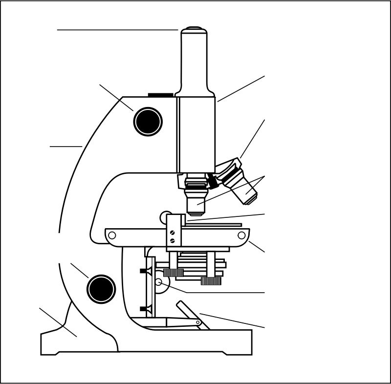
Labeling the Parts of the Microscope | Microscope World Resources Labeling the Parts of the Microscope This activity has been designed for use in homes and schools. Each microscope layout (both blank and the version with answers) are available as PDF downloads. You can view a more in-depth review of each part of the microscope here. Download the Label the Parts of the Microscope PDF printable version here.
Parts of a microscope with functions and labeled diagram - Microbe Notes Microscopes are instruments that are used in science laboratories to visualize very minute objects such as cells, and microorganisms, giving a contrasting image that is magnified. Microscopes are made up of lenses for magnification, each with its own magnification powers.
Microscope picture label Flashcards | Quizlet Microscope picture label Flashcards | Quizlet Microscope picture label STUDY Flashcards Learn Write Spell Test PLAY Match Gravity Created by kfire Terms in this set (12) Arm What is the part labelled C? Base What is the part labelled D? Body tube What is the part labelled B? Ocular lens What is the part labelled A? Illuminator
Parts of the Microscope with Labeling (also Free Printouts) Parts of the Microscope with Labeling (also Free Printouts) By Editorial Team March 7, 2022 A microscope is one of the invaluable tools in the laboratory setting. It is used to observe things that cannot be seen by the naked eye. Table of Contents 1. Eyepiece 2. Body tube/Head 3. Turret/Nose piece 4. Objective lenses 5. Knobs (fine and coarse) 6.
PDF Parts of a Microscope Printables - Homeschool Creations Label the parts of the microscope. You can use the word bank below to fill in the blanks or cut and paste the words at the bottom. Microscope Created by Jolanthe @ HomeschoolCreations.net. Parts of a eyepiece arm stageclips nosepiece focusing knobs illuminator stage objective lenses
300+ Free Microscope & Laboratory Images - Pixabay 399 Free images of Microscope Related Images: laboratory science bacteria research scientist lab biology chemistry medical Find your perfect microscope image. Free pictures to download and use in your next project.
LSM 900 with Airyscan 2 – Compact Confocal Microscope for ... In microscopy, this translates into the best contrast and resolution while maintaining minimum light exposure. LSM 900, your compact confocal microscope, provides this with components optimized to deliver the best imaging results. Get high-end confocal imaging in a small footprint. Improve any confocal experiment with LSM Plus.
PDF Label parts of the Microscope Label parts of the Microscope: . Created Date: 20150715115425Z
Looking at the Structure of Cells in the Microscope ... The phase-contrast microscope and, in a more complex way, the differential-interference-contrast microscope, exploit the interference effects produced when these two sets of waves recombine, thereby creating an image of the cell's structure . Both types of light microscopy are widely used to visualize living cells.

















Post a Comment for "44 microscope picture with labels"