40 compound microscope diagram with labels
Labelled Diagram of Compound Microscope - Biology Discussion The below mentioned article provides a labelled diagram of compound microscope. Part # 1. The Stand: The stand is made up of a heavy foot which carries a curved inclinable limb or arm bearing the body tube. The foot is generally horse shoe-shaped structure (Fig. 2) which rests on table top or any other surface on which the microscope in kept. Parts of a Compound Microscope and Their Functions Compound microscope magnification is determined by multiplying the eyepiece and objective powers. When viewed through a 5X eyepiece with a 10X objective, an item is magnified 5 x 10=50 times. The magnification is 10 x 45 = 450 times when using a 10X eyepiece and a 45X objective. How to Use the Compound Microscope
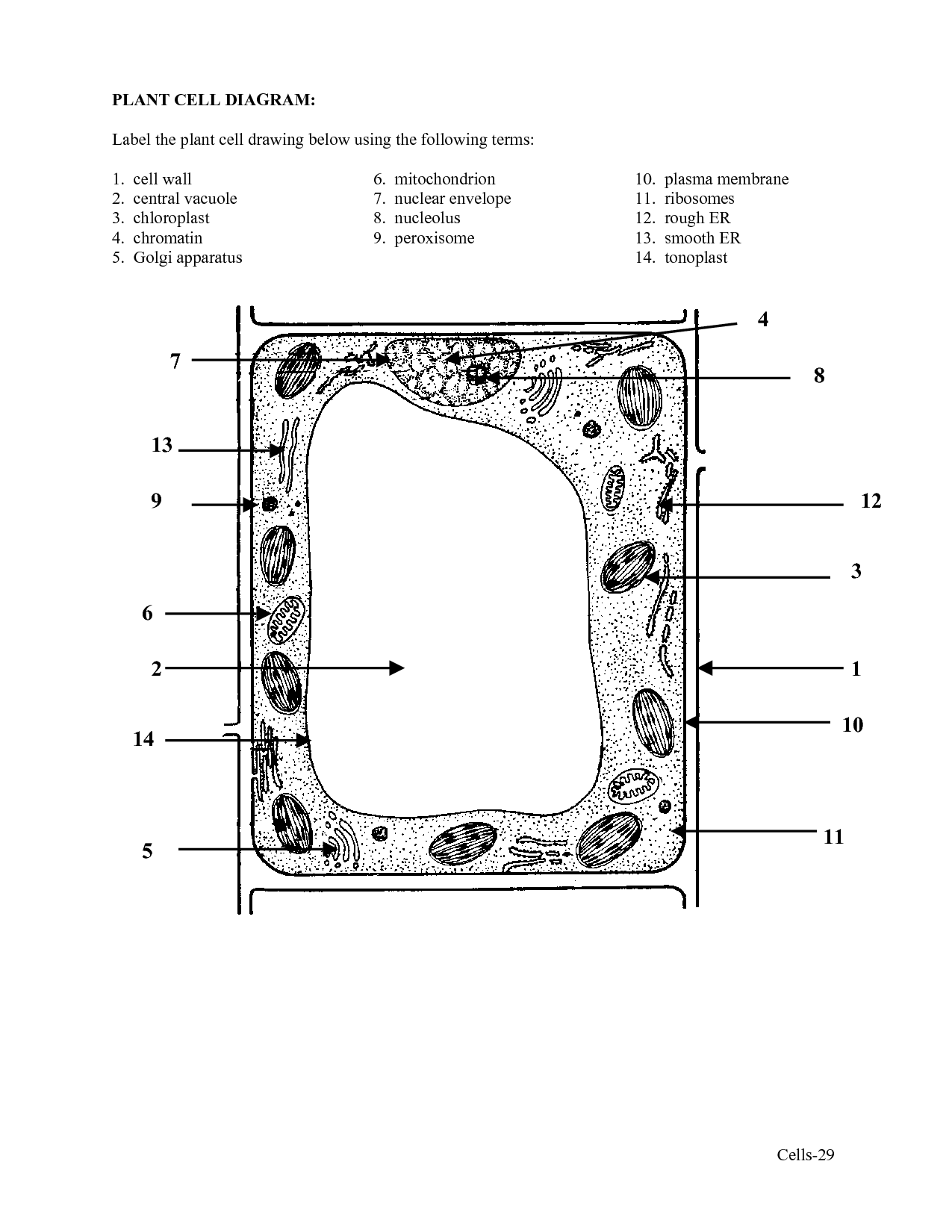
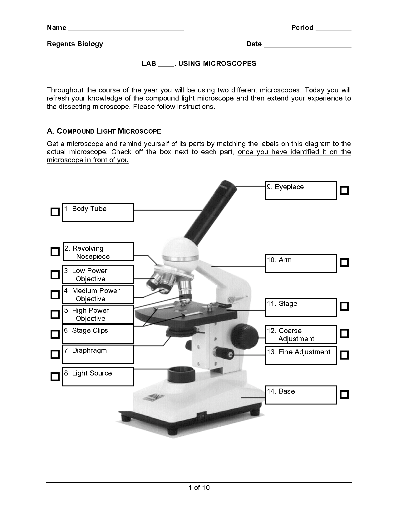
Labeling the Parts of the Microscope | Microscope activity, Science ... Description Worksheet identifying the parts of the compound light microscope. Answer key: 1. Body tube 2. Revolving nosepiece 3. Low power objective 4. Medium power objective 5. High power objective 6. Stage clips 7. Diaphragm 8. Light source 9. Eyepiece 10. Arm 11. Stage 12. Coarse adjustment knob 13. Fine adjustment knob 14. Base

Compound microscope diagram with labels
How to draw compound of Microscope easily - step by step - YouTube I will show you " How to draw compound of microscope easily - step by step "Please watch carefully and try this okay.Thanks for watching.....#microscopedrawi... Compound Microscope Parts, Diagram Definition, Application, Working ... A compound microscope is a laboratory instrument with high magnification power, which is consists of more than one lenses. Compound Microscopes are used for the study of structural details of a cell, tissue, or organ in sections. A compound microscope can magnify the image of a tiny object up to 1000. Compound Microscope Parts, Function, & Diagram - Study.com Learn the compound light microscope's parts and functions by viewing a compound microscope diagram. Also, read about the uses of a compound microscope. Updated: 11/04/2021
Compound microscope diagram with labels. Microscope Parts, Function, & Labeled Diagram - slidingmotion Microscope parts labeled diagram gives us all the information about its parts and their position in the microscope. Microscope Parts Labeled Diagram The principle of the Microscope gives you an exact reason to use it. It works on the 3 principles. Magnification Resolving Power Numerical Aperture. Parts of Microscope Head Base Arm Eyepiece Lens Microscope Types (with labeled diagrams) and Functions Compound microscope labeled diagram Compound microscope functions: It finds great application in areas of pathology, pedology, forensics etc Its greater order of magnification allows for deeper study of microbial organisms to Detect the cause of diseases Study the mineral composition in soils Microscope Parts and Functions Microscope Parts and Functions With Labeled Diagram and Functions How does a Compound Microscope Work?. Before exploring microscope parts and functions, you should probably understand that the compound light microscope is more complicated than just a microscope with more than one lens.. First, the purpose of a microscope is to magnify a small object or to magnify the fine details of a larger ... Parts of a microscope with functions and labeled diagram Figure: Diagram of parts of a microscope There are three structural parts of the microscope i.e. head, base, and arm. Head - This is also known as the body. It carries the optical parts in the upper part of the microscope. Base - It acts as microscopes support. It also carries microscopic illuminators.
Compound microscope - their parts and function - Microscopy4kids Labeled diagram of a compound microscope. Optical components of a compound microscope. The term "compound" refers to the microscope having more than one lens. Compound microscopes generate magnified images through an aligned pair of the objective lens and the ocular lens. In contrast, "simple microscopes" have only one convex lens and ... Draw a neat labelled diagram of a compound microscope and explain its ... Using sign convention, we find that O'I 1 = + v 0 and O'O = -u where v 0 is the image distance due to the objective and u is the object distance for the objective or the compound microscope. I 1 G 1 is negative and OJ is positive. To find me : The eyepiece behaves like a simple microscope. So : the magnifying power of the eye piece. ∴ m e ... Compound Microscope Parts, Functions, and Labeled Diagram Nov 18, 2020 · The individual parts of a compound microscope can vary heavily depending on the configuration & applications that the scope is being used for. Common compound microscope parts include: Compound Microscope Definitions for Labels Eyepiece (ocular lens) with or without Pointer: The part that is looked through at the top of the compound microscope. Eyepieces typically have a magnification between 5x & 30x. (i) Draw a neat labelled ray diagram of a compound microscope. Explain ... The eyepiece forms its image A'' B'' which is virtual, erect and magnified. Thus the final image A'' B'' formed by the microscope is inverted and magnified and its position is outside the objective and eyepiece towards objective lens. Magnifying power of compound microscope is. for final image at distance of distinct vision. for final image at ...
Compound Microscope Parts - Labeled Diagram and their Functions - Rs ... Labeled diagram of a compound microscope Major structural parts of a compound microscope There are three major structural parts of a compound microscope. The head includes the upper part of the microscope, which houses the most critical optical components, and the eyepiece tube of the microscope. Parts of a Compound Microscope - Labeled (with diagrams) Mar 06, 2020 · Parts of a Compound Microscope - Labeled (with diagrams) A compound microscope is known as a high-power microscope that enables you to achieve a high level of magnification. Smaller specimens can be thoroughly viewed using a compound microscope. Let us take a look at the different parts of a compound microscope and understand each key component. Image 1: The figure above is the standard image of a compound microscope. 16 Parts of a Compound Microscope: Diagrams and Video Once you have an understanding of the parts of the microscope it will be much easier to navigate around and begin observing your specimen, which is the fun part! The 16 core parts of a compound microscope are: Head (Body) Arm Base Eyepiece Eyepiece tube Objective lenses Revolving Nosepiece (Turret) Rack stop Coarse adjustment knobs Compound Microscope - Diagram (Parts labelled), Principle and Uses Feb 03, 2022 · Compound Microscope – Diagram (Parts labelled), Principle and Uses As the name suggests, a compound microscope uses a combination of lenses coupled with an artificial light source to magnify an object at various zoom levels to study the object. A compound microscope: Is used to view samples that are not visible to the naked eye

Microscope With Labels Clip Art at Clker.com - vector clip art online ...
Compound Microscope Labeled Diagram | Quizlet QUESTION. The total magnification of a specimen being viewed with a 10X ocular lens and a 40X objective lens is. 15 answers. QUESTION. a mosquito beats its wings up and down 600 times per second, which you hear as a very annoying 600 Hz sound. if the air outside is 20 C, how far would a sound wave travel between wing beats. 2 answers.

31 Label Of A Microscope - Label Design Ideas 2020
Diagram of a Compound Microscope - Biology Discussion The size of objects viewed under the compound microscope can be accurately determined using a micrometer. The latter consists of two scales, the eyepiece scale, (also called 'graticule' or 'ocular') and the stage micrometer scale. The eyepiece scale is calibrated with the help of stage micrometer and the former is then used for measurements.

compound light microscope labeled 28125 - Made By Creative Label
Label Parts Of A Compound Microscope Teaching Resources | TpT This is a set of 3 tiered readings. Students will read a passage about the how to use a compound light microscope. Students will use textual evidence to answer questions and label the different parts of the microscope. It also allows students to gain prior knowledge about the compound microscope. Version A provides the most support for students.
16 Best Images of Simple Microscope Labeling Worksheet - Compound Light ...
Label the microscope — Science Learning Hub Label the microscope Add to collection Use this interactive to identify and label the main parts of a microscope. Drag and drop the text labels onto the microscope diagram. eye piece lens coarse focus adjustment high-power objective diaphragm or iris base fine focus adjustment light source stage Download Exercise Tweet
Microscope With Labels Clip Art at Clker.com - vector clip art online ...
PDF COMPOUND LIGHT MICROSCOPE LAB - Springfield Public Schools COMPOUND(LIGHT(MICROSCOPE(LAB((Follow(written(andoral(instructions.((((A.((Label(the(parts(of(the(compound(microscope.(Be(able(to(label(a(blank(diagram
8 Best Images of Using A Microscope Worksheet - Compound Microscope ...
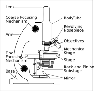
Parts of the Compound Microscope - HCC Learning Web Parts of the Compound Microscope Use Figure 2 as a guide to locate the major parts of the compound microscope. a. Base: The bottom, flat part that supports the microscope. b. Arm: The straight or curved vertical part that connects the base to the upper portion. c. Body Tube: Extends from the arm and contains the ocular lens and the rotating

Microscope And Its Types | Medical and Biological Types
A Study of the Microscope and its Functions With a Labeled Diagram To better understand the structure and function of a microscope, we need to take a look at the labeled microscope diagrams of the compound and electron microscope. These diagrams clearly explain the functioning of the microscopes along with their respective parts. Man's curiosity has led to great inventions. The microscope is one of them.

Diagrams of Microscope | 101 Diagrams
Compound Microscope: Definition, Diagram, Parts, Uses, Working ... - BYJUS Compound microscope is a type of optical microscope that is used for obtaining a high-resolution image. There are more than two lenses in a compound microscope. Learn about the working principle, parts and uses of a compound microscope along with a labeled diagram here.

Anatomy and Physiology I Coursework: Microscope A+P
Parts of Stereo Microscope (Dissecting microscope) - labeled diagram ... Labeled part diagram of a stereo microscope Major structural parts of a stereo microscope There are three major structural parts of a stereo microscope. The viewing Head includes the upper part of the microscope, which houses the most critical optical components, including the eyepiece, objective lens, and light source of the microscope.
Post a Comment for "40 compound microscope diagram with labels"