43 how to label nmr spectra
13C Carbon NMR Spectroscopy - Chemistry Steps In general, when you start analyzing a 13 C NMR, split the spectrum in two parts by drawing a line at 100 ppm; below this value you have the saturated functional groups, and beyond that is the unstructured region. Saturated carbon atoms connected to electronegative heteroatoms give signal from 30-90 ppm. PDF NMR of Proteins - University at Buffalo • simple 152D 1H, N correlation NMR spectra of proteins • reduce complex spectra to simple ones based on isotope editing • reduce/eliminate spectral overlap/spectral degeneracy • correlate amide 1H-15N pairs • this spectrum demonstrates 1). that you can express your protein, 2). That you can isotopically label your protein, 3). That your
A Step-By-Step Guide to 1D and 2D NMR Interpretation Now we have a fully assigned ¹H-NMR spectrum! This spectrum will help us assign our carbons using HSQC and HMBC NMR spectroscopy. Step 3: ¹³C-NMR Carbon NMR is a necessary step in full structural characterization. However, ¹³C-NMR alone does not provide enough information to assign the carbons in the molecule.

How to label nmr spectra
PDF NMR Guidelines for ACS Journals 1.1 The compound must be clearly identified, for example in a header at the beginning of a) the synthetic procedure or b) the summary of spectroscopic data. 1.2 List the nucleus being measured, any nucleus being broadband decoupled, the - solvent used (formula preferred, e.g. C 6 D 6 over benzene- d 6 NMR Spectroscopy - Michigan State University The following features lead to the nmr phenomenon: 1. A spinning charge generates a magnetic field, as shown by the animation on the right. The resulting spin-magnet has a magnetic moment ( μ) proportional to the spin. 2. In the presence of an external magnetic field ( B0 ), two spin states exist, +1/2 and -1/2. Simulate and predict NMR spectra Predict COSY spectra. Predict HSQC / HMBC spectra. Simulated second order effect in 1 H NMR spectra. Recreate (resurect) 1D NMR spectra from experimental in-line assignment. 1 H NMR prediction was possible thanks to the tool of the FCT-Universidade NOVA de Lisboa developped by Yuri Binev and Joao Aires-de-Sousa. Y.
How to label nmr spectra. PDF Examination of Proton NMR Spectra considerably easier than analysis of proton spectra. In most cases the analysis of a C-13 spectrum involves noting the number of signals and their chemical shift. Each carbon gives rise to a signal, but if two or more carbons are equivalent, their signals will exactly overlap. As in proton NMR chemical shift may suggest the type of Nuclear magnetic resonance (NMR) spectroscopy: Hydrogen Hydrogen NMR. NMR is particularly useful in the identification of the positions of hydrogen atoms ( 1 H) in molecules. The NMR spectrum of ethyl benzene, C 6 H 5 CH 2 CH 3, is shown below.The frequencies correspond to the absorption of energy by 1 H nuclei, which are protons. Notice that there are three major peaks of differing heights. How to reference 1D and 2D NMR spectra? … Part 3 - ACD/Labs When it comes to referencing 2D NMR data, use the 1D NMR data collected at the same temperature and solvent. The 1H NMR spectra below are for the same compound quinine prepared with different deuterated solvents. The shifts in the peaks are a result of the different effects of the solute-to-solvent interactions. When analyzing 2D NMR data for ... How to interpret NMR spectra - University of Texas at Austin 1.2 An NMR spectrum is a plot of absorbance versus frequency. 1.2A To make different spectra directly comparable, a standard is used for all NMR spectra. For 1H NMR spectra, the standard is called tetramethylsilane (TMS) and a small amount of TMS is usually added to any 1H NMR sample.
NMR - Interpretation - Chemistry LibreTexts NMR - Interpretation. Nuclear Magnetic Resonance (NMR) interpretation plays a pivotal role in molecular identifications. As interpreting NMR spectra, the structure of an unknown compound, as well as known structures, can be assigned by several factors such as chemical shift, spin multiplicity, coupling constants, and integration. interpreting C-13 NMR spectra - chemguide Using the simplified table of chemical shifts above, work out the structure of the compound with the following C-13 NMR spectrum. Its molecular formula is C 4 H 6 O 2. Let's sort out what we've got. There are four peaks and four carbons. No two carbons are in exactly the same environment. 6.7 ¹H NMR Spectra and Interpretation (Part II) With the structure of a compound given, we can apply all the knowledge about 1 H NMR to assign the signals in the spectrum, that is to identify a certain signal comes from which hydrogen (s). Examples Match the 1 H NMR spectrum below to its corresponding compound, and assign all of the signals. NMR Spectrum of Phenacetin | Thermo Fisher Scientific - US Phenacetin (C 10 H 13 NO 2 ), N- (4-Ethoxyphenyl)acetamide, is a drug that saw widespread application for nearly a century in many over-the-counter medications as a remedy for pain and fever. The 82 MHz spectrum of a 6% (w/w; 0.5 M) solution of phenacetin in CDCl 3 acquired using the Thermo Scientific picoSpin 80 NMR spectrometer.
NMR Spectroscopy Principles, Interpreting an NMR Spectrum and Common ... By convention, the chemical shift scale in an NMR spectrum is represented from right to left. As described above, the zero value is established using a standard compound whose carbon and hydrogen atoms are strongly shielded and hence, their signals appear in the furthest upfield region (as seen in Figure 7). high resolution nuclear magnetic resonance (nmr) spectra In a high resolution spectrum, you find that many of what looked like single peaks in the low resolution spectrum are split into clusters of peaks. For A'level purposes, you will only need to consider these possibilities: 1 peak. a singlet. 2 peaks in the cluster. a doublet. 3 peaks in the cluster. a triplet. How To Label NMR Spectra - YouTube About Press Copyright Contact us Creators Advertise Developers Terms Privacy Policy & Safety How YouTube works Test new features Press Copyright Contact us Creators ... NMR- drawing and labelling spectra. - The Student Room #1 Hi, I've been given the molecule PF2H and I have to sketch the phosporous, flourine and hydrogen NMR spectra. Then label the peaks and coupling constants. I think I've worked out the shape to be trigonal bipyramidal but I'm a bit confused about drawing the spectra.
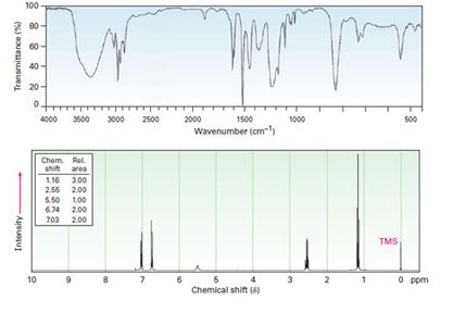
How to Identify Molecular Fragments from NMR and IR Spectra The IR and NMR spectra for a compound with molecular formula C 5 H 10 O Carbonyl groups are found in aldehydes, ketones, esters, and carboxylic acids. Here, however, esters and carboxylic acids can be eliminated because the molecular formula shows only a single oxygen.
PDF Analysis of H Nmr Spectra show two signals in the proton NMR spectrum. There is a direct relationship between the symmetry of the molecule and the number of proton NMR signals it exhibits. The following example illustrates this point dramatically. The cyclohexane molecule exhibits only one signal at room temperature. The mere introduction of a methyl group changes the
Assigning a 1H NMR spectrum - YouTube In this screencast, Andrew Parsons walks you through the process of assigning a 1H NMR spectrum.
6.6 ¹H NMR Spectra and Interpretation (Part I) Generally, the information about the structure of molecule can be obtained from four aspects of a typical 1 H NMR spectrum: Chemical equivalent and non-equivalent protons (total number of signals) Chemical shift Integration Signal splitting 6.6.1 Chemical Equivalent and Non-Equivalent Protons
NMR Spectrum of Aspirin | Thermo Fisher Scientific - US The 1 H NMR spectrum of a 4% (w/w; 330 mM) solution of aspirin in chloroform-D was measured at 82 MHz using the Thermo Scientific picoSpin 80 NMR spectrometer. Aspirin contains aliphatic, aromatic and carboxylic acid protons that span a wide range of the 1H spectrum, and signal integration reveals a 3:4:1 intensity distribution, respectively ...
Isotopic Labeling for NMR Spectroscopy of Biological Solids The simplest and most cost-effective biosynthetic labeling method for protein solid-state NMR is to uniformly label all carbon and nitrogen atoms with 13 C and 15 N. In this way, a single protein sample can in principle provide all the structural constraints - dihedral angles and distances - about the protein.
PDF How to Plot and Print NMR Spectrum - Swarthmore College FIRST, CLICK THE SIDE OF THE REPORT IMAGE WHERE THERE IS NO OBJECT. THE LEFT SIDE TOOLBAR WILL DISPLAY THE OPTION TO ADD A NMR SPECTRUM. USING THE MOUSE, LEFT CLICK-HOLD AND DRAG TO FORM A BOX WHERE THE INSET WILL BE. CLICKING INSIDE THE INSET SPECTRUM YOU CAN CHANGE THE SCALE, SIZE, X AND Y-AXIS LIMITS LIKE THE MAIN SPECTRUM.
Deciphering 1H NMR Spectra - Organic Chemistry Help Once a NMR spectrograph is recorded, 4 pieces of information can be determined from the data as long as the chemical formula of the compound is known. To illustrate the points, we will consider the following 1H-NMR spectrum of the C 5 H 10 O. Signal Count - Number of unique hydrogens This is the easiest to interpret.
How to Interpret Proton NMR Spectra - theSpectroscopy Now that we have had an introduction to key aspects of 1 H NMR spectra (chemical shift, peak area, and signal splitting), we can start to apply 1 H NMR spectroscopy to elucidating the structure of unknown compounds. The following steps summarize the process:


Post a Comment for "43 how to label nmr spectra"