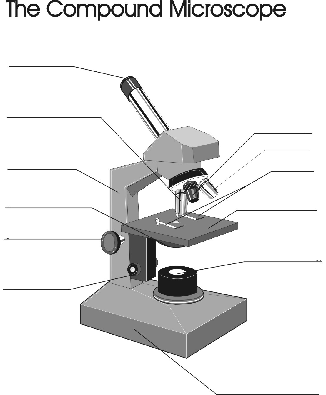
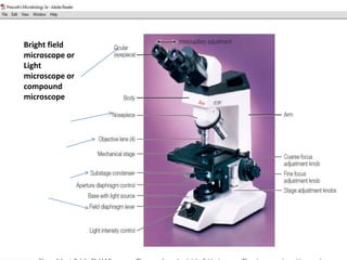
42 compound microscope unlabeled
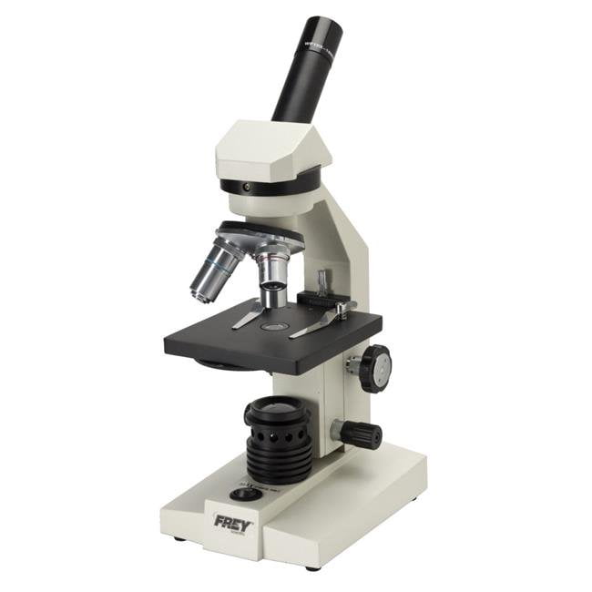
Compound Microscope Parts Head/Body houses the optical parts in the upper part of the microscope. Base of the microscope supports the microscope and houses the illuminator. Arm connects to the base and supports the microscope head. It is also used to carry the microscope. When carrying a compound microscope always take care to lift it by both the arm and base ... Microscope Images Labeled | Virtual Anatomy Lab VAL - ncccval Body cavities, planes, and regions. Body Images Labeled. Body Images Unlabeled. Histology. Epithelium Images Labeled. Epithelium Images Unlabeled. Connective Tissue Images Labeled. Connective Tissue Images Unlabeled. Microscope.
Compound Microscope Parts - Labeled Diagram and their Functions The term "compound" refers to the microscope having more than one lens. Basically, compound microscopes generate magnified images through an aligned pair of the objective lens and the ocular lens. In contrast, "simple microscopes" have only one convex lens and function more like glass magnifiers. [In this figure] Two "antique" microscopes played significant roles in the history of biology.

Compound microscope unlabeled
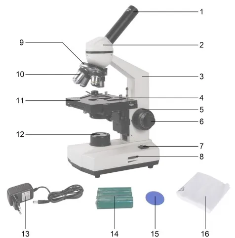
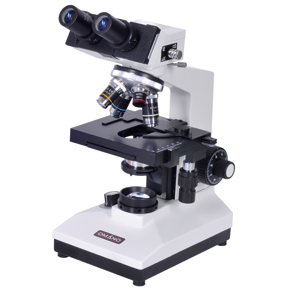
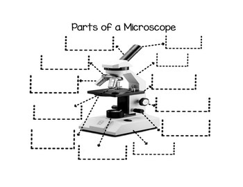
Microscope Parts and Functions Body tube (Head): The body tube connects the eyepiece to the objective lenses. Arm: The arm connects the body tube to the base of the microscope. Coarse adjustment: Brings the specimen into general focus. Fine adjustment: Fine tunes the focus and increases the detail of the specimen. Nosepiece: A rotating turret that houses the objective lenses. Compound Microscope Parts, Functions, and Labeled Diagram Compound Microscope Definitions for Labels. Eyepiece (ocular lens) with or without Pointer: The part that is looked through at the top of the compound microscope. Eyepieces typically have a magnification between 5x & 30x. Monocular or Binocular Head: Structural support that holds & connects the eyepieces to the objective lenses. Amazon.com: AmScope M620C-E1 Digital Compound Monocular Microscope ... This item: AmScope M620C-E1 Digital Compound Monocular Microscope, WF10x and WF25x Eyepieces, 40x-2500x Magnification, Brightfield, LED Illumination, Abbe Condenser, Mechanical Stage, 110V, Includes 1.3MP Camera and Software. $281.99. Only 1 left in stock (more on the way). Ships from and sold by Amazon.com.
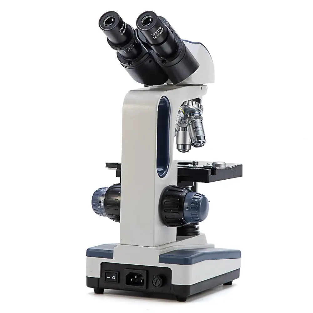
Compound microscope unlabeled. Digital Binocular Compound Microscope | Microscope Central This digital binocular compound microscope is perfect for veterinary practices, routine labs, or education. This all-in-one digital binocular compound microscope captures 16.0 M.P. images and videos up to 4 M.P. For quality digital imaging microscopy, the integrated computer runs Windows 10 with a hard drive offering internal storage, ethernet, and WiFi access, and an HDMI output to connect to a larger device. labeling a microscope worksheet microscope labeled unlabeled timvandevall clearboth. 17 Best Images Of Microscope Labeling Worksheet - Microscope Parts Quiz ... . microscope parts compound worksheet diagram printable worksheets quiz grade biology labeling science answers sketch labeled tests blank microscopio light label. 17 Best Images Of Microscope Labeling ... Compound Microscope Labeled Diagram | Quizlet Part that supports the microscope. Stage. Supports the slide or specimen. Coarse adjustment Knob. sed to focus when using the low power objective lenses. Fine Adjustment Knob. Used to focus the image on high power to view image in more detail. Revolving nose piece. The revolving piece on which the lenses are attached. Positive and Unlabeled Materials Machine Learning Image by author. We calculated a bunch of properties for all the materials we were interested in, built and trained a positive and unlabeled machine learning model to recognize what is special about the synthesized materials, and then predicted which new materials should be synthesizable. Our model learned to use some information that we already know is a good indicator of synthesizability ...
Parts of the Microscope with Labeling (also Free Printouts) 5. Knobs (fine and coarse) By adjusting the knob, you can adjust the focus of the microscope. The majority of the microscope models today have the knobs mounted on the same part of the device. Image 5: The circled parts of the microscope are the fine and coarse adjustment knobs. Picture Source: bp.blogspot.com. How to see a plant cell under a compound microscope - Quora Answer (1 of 2): There four focus level in compound microscope 4X,10X,40X and 100X Just place your prepared slide of plant between light and slide stand and focus on 40X or 100X you can easily see plant cells under microscope How Much Does a Microscope Cost? (With 20 Microscope Prices) It is very important to adjust the condenser to the optimal numerical aperture for the objective that you are using, and it is difficult to get it just right on an unlabeled condenser. Most condensers that come with lower priced microscopes have unlabeled standard 1.25 numerical aperture Abbe condensers. Microscope Types (with labeled diagrams) and Functions Has a higher level of magnification - Typically up to 2000x. Is used in hospitals and forensic labs by scientists, biologists and researchers to study micro organisms. Compound microscope labeled diagram. Compound microscope functions: It finds great application in areas of pathology, pedology, forensics etc.
Lab 1 Microscopes and Cells.docx - Lab 1 Microscopes and... Place unlabeled microscope diagrams in lab notebook and neatly label. Viewing letter "e" slide under the Dissecting Microscope 1. Place the slide on the stage of the dissecting microscope. 2. Use trans illumination to illuminate specimen. 3. Focus the microscope at LOWEST magnification and draw/take a picture of the image seen. 4. Morphology Analysis of Unlabeled Red Blood Cells based ... - ResearchGate The approach consists of using a compact, custom-built microscope to record large field-of-view, bright-field and fluorescence images of samples that are stained with a single dye, and using ... Microscope Diagram Labeled, Unlabeled and Blank | Parts of a Microscope ... Microscope Diagram Labeled, Unlabeled and Blank | Parts of a Microscope. Print a microscope diagram, microscope worksheet, or practice microscope quiz in order to learn all the parts of a microscope. ... Basic steps for focusing a compound light microscope. Suburban Science. Biology: Cells. Vet Tech School. Vet Tech Student. Vet Medicine ... Compound Microscope- Definition, Labeled Diagram, Principle, Parts, Uses A compound microscope is of great use in pathology labs so as to identify diseases. Various crime cases are detected and solved by drawing out human cells and examining them under the microscope in forensic laboratories. The presence or absence of minerals and the presence of metals can be identified using compound microscopes.
OMAX Microscope Monocular Compound Microscope 40x~800x with ...
PDF AN INTRODUCTION TO THE COMPOUND MICROSCOPE - Rowan University microscope in an upright position using both hands. **When carrying the microscope, place one hand on the base and the other hand around the arm. **DO NOT PLACE THE MICROSCOPE IN AN UPSIDE DOWN POSITION. PIECES WILL FALL OUT. **Keep microscope away from the edge of the bench, particularly when not in use. **Make sure power cords are out of the way.

Microscope Components - Science Quiz
Compound Microscope: Parts of Compound Microscope - BYJUS Foot or base. It is a U-shaped structure and supports the entire weight of the compound microscope. 2. Pillar. It is a vertical projection. This stands by resting on the base and supports the stage. 3. Arm. The entire microscope is handled by a strong and curved structure known as the arm.

Living Environment Course
The Compound Light Microscope - Miami University The microscope pictured above is referred to as a compound light microscope. The term light refers to the method by which light transmits the image to your eye. C ompound deals with the microscope having more than one lens. Microscope is the combination of two words; "micro" meaning small and "scope" meaning view.
(159).jpg)
Microscope Quiz: How Much You Know About Microscope Parts And ...
Oops! | Flickr We're having some trouble displaying this photo at the moment. Please try again.

LAB 1: HISTOLOGY Diagram | Quizlet
Microscope Diagram Labeled, Unlabeled and Blank - Pinterest When students map a microscope, they assign colors and/or patterns to each part they need to identify. They fill in the key and the corresponding part. This file includes two types of microscope maps: 1. a microscope with the twelve parts listed along the side 2. a microscope with arrows that allows the student to…
Frey Scientific 528600 LED Illumination National Optical ...
A Study of the Microscope and its Functions With a Labeled Diagram The compound microscope uses light for illumination. Some compound microscopes make use of natural light, whereas others have an illuminator attached to the base. The specimen is placed on the stage and observed through different lenses of the microscope, which have varying magnification powers. Compound Microscope Parts and Functions

Compound Microscope Parts
Labeling the Parts of the Microscope | Microscope World Resources Microscope World explains the parts of the microscope, including a printable worksheet for schools and home. Need Asssistance? 800-942-0528. Microscope Blog. Resource Library . Login Request Quote. 0. 0. Industrial Stereo Microscopes. Digital; Lighted Stand; Plain Stand + External Light; Articulated Arm;

Microscope With Labels Clip Art at Clker.com - vector clip ...
BIO201-Microscope This page last updated 2 September 2019 by Udo M. Savalli ()Images and text © Udo M. Savalli. All rights reserved.

Optical microscope - 037 - Breukhoven - laboratory ...
the compound microscope worksheet microscope diagram blank labeled worksheet labeling parts compound worksheeto light heart via unlabeled quiz. Compound Microscope Labeling Worksheet - WorksSheet List atehnyerbl0g.blogspot.com. microscope labeling. Compound Microscope Parts Worksheet justiceforallcitizens.com. answers optical.
40x-1600x Led Lab Monocular Compound Microscope With 3d-stage ...
Label the microscope — Science Learning Hub All microscopes share features in common. In this interactive, you can label the different parts of a microscope. Use this with the Microscope parts activity to help students identify and label the main parts of a microscope and then describe their functions.. Drag and drop the text labels onto the microscope diagram. If you want to redo an answer, click on the box and the answer will go back ...

Amazon.co.uk Best Sellers: The most popular items in Compound ...
Microscope Labeling Game - PurposeGames.com This is an online quiz called Microscope Labeling Game. There is a printable worksheet available for download here so you can take the quiz with pen and paper. This quiz has tags. Click on the tags below to find other quizzes on the same subject. Science. microsope. Your Skills & Rank. Total Points. 0. Get started! Today's Rank--0.

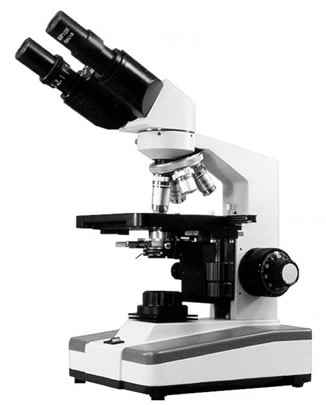
OMAX Microscope 2.0MP Digital Compound Siedentopf Binocular ...
Amazon.com: AmScope M620C-E1 Digital Compound Monocular Microscope ... This item: AmScope M620C-E1 Digital Compound Monocular Microscope, WF10x and WF25x Eyepieces, 40x-2500x Magnification, Brightfield, LED Illumination, Abbe Condenser, Mechanical Stage, 110V, Includes 1.3MP Camera and Software. $281.99. Only 1 left in stock (more on the way). Ships from and sold by Amazon.com.

Buy National Optical 40X-1000X Compound Microscope Set with ...
Compound Microscope Parts, Functions, and Labeled Diagram Compound Microscope Definitions for Labels. Eyepiece (ocular lens) with or without Pointer: The part that is looked through at the top of the compound microscope. Eyepieces typically have a magnification between 5x & 30x. Monocular or Binocular Head: Structural support that holds & connects the eyepieces to the objective lenses.

Microscope Labeling
Microscope Parts and Functions Body tube (Head): The body tube connects the eyepiece to the objective lenses. Arm: The arm connects the body tube to the base of the microscope. Coarse adjustment: Brings the specimen into general focus. Fine adjustment: Fine tunes the focus and increases the detail of the specimen. Nosepiece: A rotating turret that houses the objective lenses.

Compound Microscope Parts, Functions, and Labeled Diagram ...

Label Microscope Diagram - EnchantedLearning.com

parts of the microscope

Microscope Diagram Labeled, Unlabeled and Blank | Parts of a ...

AmScope - 40X-2500X LED Digital Monocular Compound Microscope w 3D Stage +1.3MP USB Imager

Compound Microscope Parts, Functions, and Labeled Diagram ...

ilk Birmanya üç 2000x mikroskop - tridigiwet.com
Remix of "The Compound Microscope"

Compound microscope - their parts and function - Microscopy4kids

Binocular Compound Light Microscope Diagram Quiz

Compound microscope - their parts and function - Microscopy4kids

Amazon.com: AmScope B690B Siedentopf Binocular Compound ...

Swift Trinocular Compound Microscope SW350T,40X-2500X ...
Biological Microscope Kit For Vet Clinical Lab College ...
Parts of a Compound Microscope - Labelled diagram
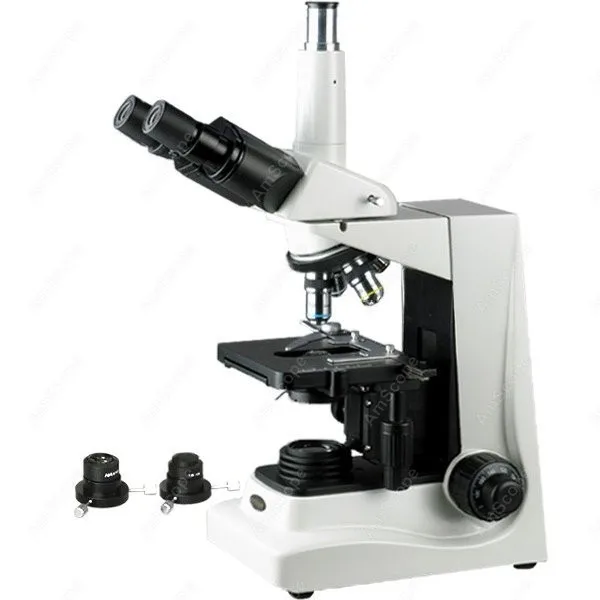
Darkfield Brightfield Trinocular Microscope--AmScope Supplies Darkfield Brightfield Trinocular Compound Microscope 40X-1600X

Biology 30 Compound Microscope Diagram | Quizlet

Microscope Use Lab Purpose: To learn the parts and how to use ...
Microscope | Analytical Wiki | Fandom

OMAX Microscope 1600X Oil Darkfield & Brightfield Siedentopf ...

Celestron CM2000CF - Compound Microscope
Microscopy
Muscle Anatomy Study Cards Flashcards | Chegg.com

Label the microscope — Science Learning Hub

40X-2000X Student Biological Microscope Top Bottom LED Illuminated Microscope | eBay

Compound Light Microscope Diagram | Quizlet
Microscope Foldable Teaching Resources | Teachers Pay Teachers

Introduction to the Compound Light Microscope Chuck Hesbacker ...
Post a Comment for "42 compound microscope unlabeled"