45 spirogyra microscope labeled
Spirogyra Under Microscope Labeled : Vegetative Spirogyra Prepared ... Spirogyra Under Microscope Labeled : Vegetative Spirogyra Prepared Microscope Slide 75x25mm Eisco Labs - Besides, the filaments are also surrounded by mucilage that holds the filaments together to form clumps in water.. The beauty of spirogyra is most prominent under the microscope, where helices of. The triatomine bug thrives under poor ... Spirogyra Vector Illustration. Labeled Educational Green Algae ... Spirogyra Vector Illustration. Labeled Educational Green Algae Structure. Stock Vector - Illustration of isolated, biology: 168786837 Stand with Ukraine! 5% of our sales go to NGOs supporting Ukrainian causes and war refugees. More about Dreamstime Giving Fund. Our Ukrainian photographers and illustrators. Get 15 images free trial
Spirogyra Labelled Diagram Spirogyra (common names include water silk, mermaid's tresses, and blanket weed) is a genus of filamentous charophyte green algae of the order Zygnematales, named for the helical or spiral arrangement of the chloroplasts that is characteristic of the genus. Draw a labelled diagram of Spirogyra. 51 Differentiate between flying lizard and bird.

Spirogyra microscope labeled
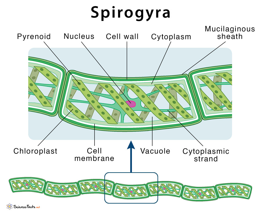
Spirogyra_diagram_labeled | Spirogyra diagram, Teaching biology, Middle ... Spirogyra_diagram_labeled Find this Pin and more on Algae by Aerobe. More like this Biology Projects Biology Art Biology Lessons Cell Biology School Projects Science Models Single Celled Wooden Main Door Design Green Algae C (914) 456-6009 Dna Microscopic Algae Microscopic Images Science Biology Flora Protists Bio Art Plant Cell Labeled Diagram of Spirogyra - QS Study Spirogyra is a sophisticated, filamentous green alga, found in freshwater represented by about 300 species. It is also identified as pond silk, as its fiber burnishes like silk due to the occurrence of mucilage. The vegetative body structure of spirogyra. A) External features. The vegetative body of Spirogyra is unbranched and filamentous. Spirogyra: Structure, Diagram, Fragmentation, Sexual Reproduction - BYJUS Spirogyra are free-floating green algae present in freshwater habitats such as ponds, lakes, etc. Spirogyra are commonly known as "water silk or pond silk". They have a filamentous and unbranched vegetative structure. There are around 400 species of Spirogyra found.
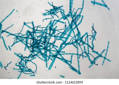
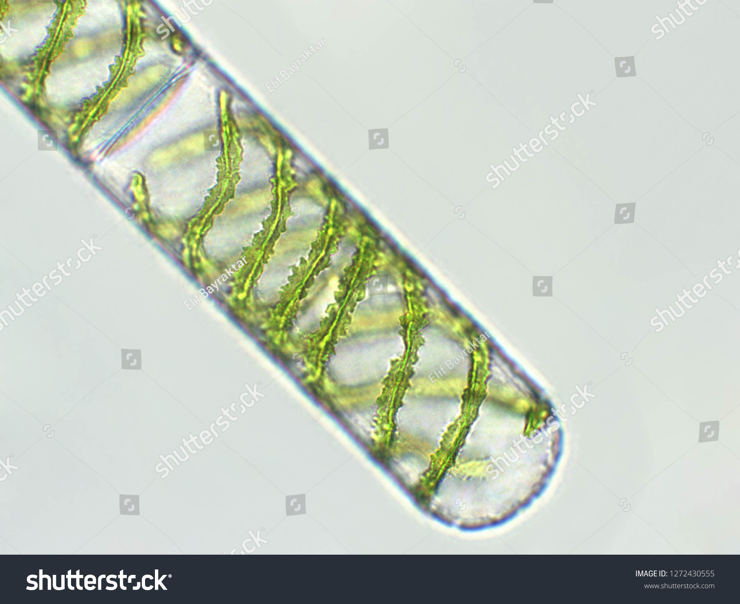
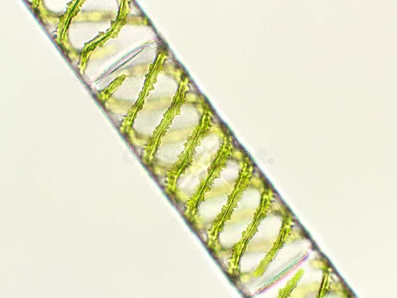
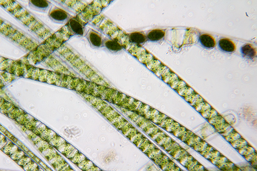
Spirogyra microscope labeled. spirogyra | Definition, Structure, Reproduction, & Facts spirogyra, (genus Spirogyra), any member of a genus of some 400 species of free-floating green algae (division Chlorophyta) found in freshwater environments around the world. Named for their beautiful spiral chloroplasts, spirogyras are filamentous algae that consist of thin unbranched chains of cylindrical cells. They can form masses that float near the surface of streams and ponds, buoyed by ... Under The Microscope: Paramecium - Office for Science and Society Paramecium are single-celled organisms that belong to the Ciliophora phylum. Members of this group are characterized by having cilia, or little hair-like structures covering their surface. Once called "slipper animalcules" due to their oblong shape, Paramecium live in a variety of watery environments, both fresh and salt, although they are ... What is Spirogyra? (Characteristics ... - Microscope Clarity Spirogyras are found in freshwater environments like shallow ponds, ditches, and at the edges of lakes. They are generally free-floating and can be found in large mats of other Spirogyra. Spirogyra are unique in that they are short-lived and are most abundant during periods of wet weather. They are known to dry up very quickly. Spirogyra - Wikipedia Spirogyra (common names include water silk, mermaid's tresses, and blanket weed) is a filamentous charophyte green alga of the order Zygnematales, named for the helical or spiral arrangement of the chloroplasts that is characteristic of the genus. It is commonly found in freshwater habitats, and there are more than 400 species of Spirogyra in the world.
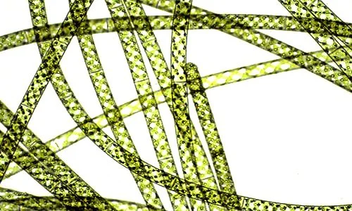
2 place the slide labeled spirogyra on the microscope Place the slide labeled Spirogyra on the microscope and view the slide under low, medium, and high powers. Note: Use the lens paper as necessary to wipe the slide. 1. Use the background section, a textbook, and/or an Internet source to determine if the Spirogyra is a protist, plant, animal, or bacteria. Record in Data Table 2. 2. Onion Plant Cell Under Microscope Labeled - Ismael Dauila The cell is in prophase of mitosis, with distinct chromosomes plant cell under microscope labeled. Only consider those cells that are filled with the red pigment. Source: faculty.kutztown.edu. Microscopic view of an onion skin showing several rectangular cells, each with a small, spherical nucleus (red arrow). Spirogyra Under The Microscope - YouTube Spirogyra is a filamentous green algae found in freshwater environments. It is often found as green clumps, although each strand is microscopic. Spirogyra gets its name from the spiral pattern of... Hilaire Simon: Euglena Cell Under Microscope Labeled - Euglena ... Euglena spirogyra plant cell, microorganisms, microbiology, science, . A labeled euglena diagram showing the structural features visible in . Other species, such as euglena viridis and . Euglena species are protozoa that are capable of photosynthesising (like plants) and consuming other organisms (like predatory animals).
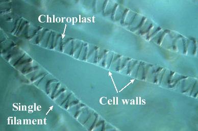
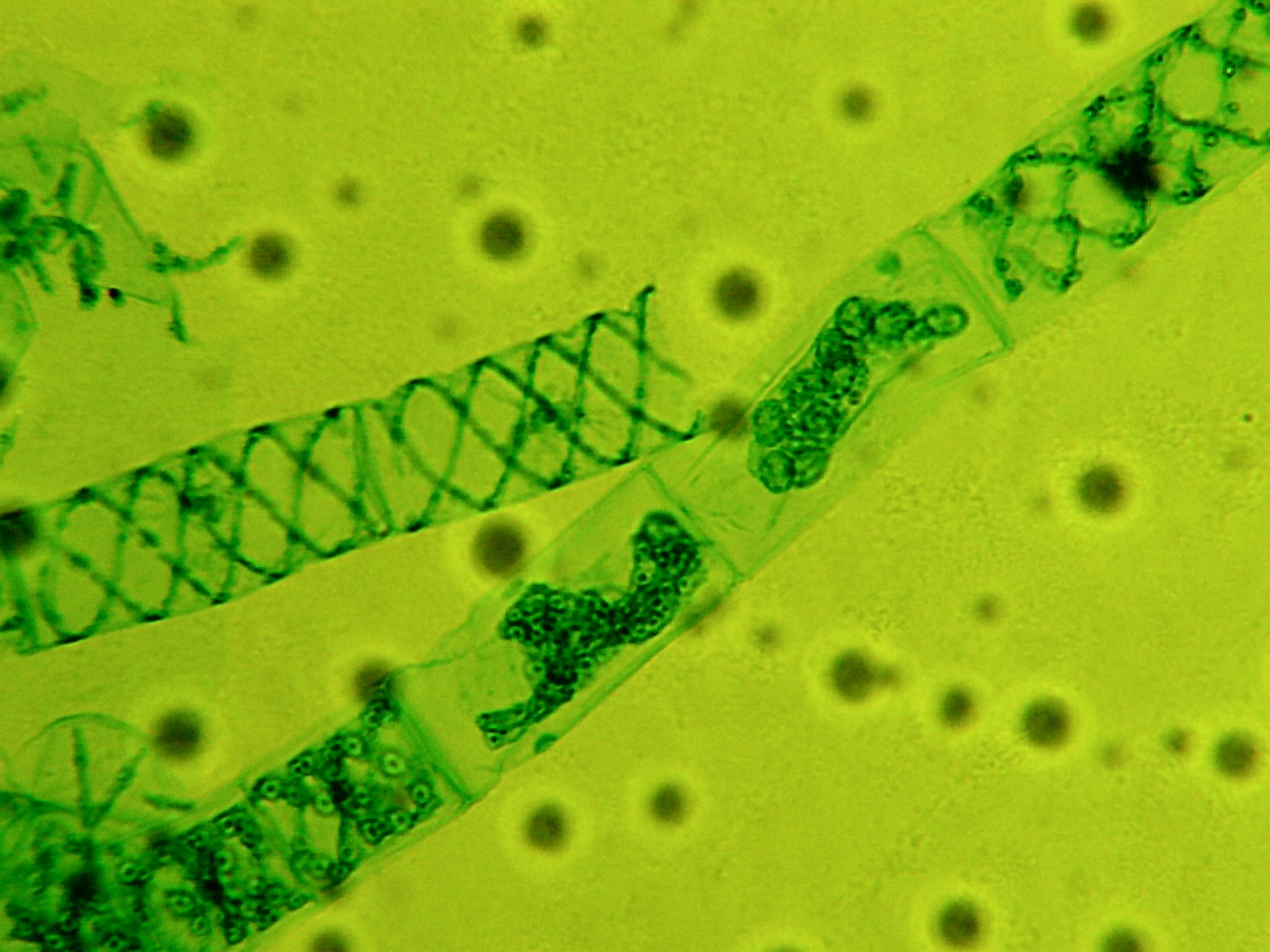
Microscope Imaging Station. Gallery. - Exploratorium Some plant cells have organelles called chloroplasts that make them green and able to capture energy from light. Rigid walls typically made of cellulose surround plant cells. Video: Spirogyra, structures labeled This algae grows in long, hairlike strands in freshwater ponds. The helical green structures inside the cell walls are chloroplasts. Worksheet_Basic_Microscopy.html - Basic Microscopy_Essay1_Labeled ... Basic Microscopy_Essay1_Labeled Amoeba Image EXPERIMENT 2: Upload your labeled Amoeba image here. 2 Basic Microscopy_Essay2_Labeled Spirogyra Image EXPERIMENT 2: Upload your labeled Spirogyra image here. 3 Basic Microscopy_Essay3_Describe Spirogyra Cells EXPERIMENT 2: Describe the shape of the Spirogyra cells. Which cellular structure gives rise to this shape? 4 Basic Microscopy_Essay4_Labeled ... spirogyra Flashcards and Study Sets | Quizlet Spirogyra -practical 1. How does spirogyra move? What is spirogyra's niche? Where does spirogyra live? moves towards the surface of water closer to light using cilia. producer. freshwater, ponds, ditches, and clean water w/ lots of nutrien…. 17 Terms. Spirogyra - Introduction, Structure and Reproduction - VEDANTU Spirogyra is a kind of algae that is studied in chapters that are based on plant reproduction. In many parts of the world, spirogyra has multiple names such as mermaid's tresses, pond scum, water-silk. The common name for SpiroGyra is green algae. SpiroGyra is a member of a genus of about 400 species which are free-floating green algae ...
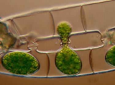
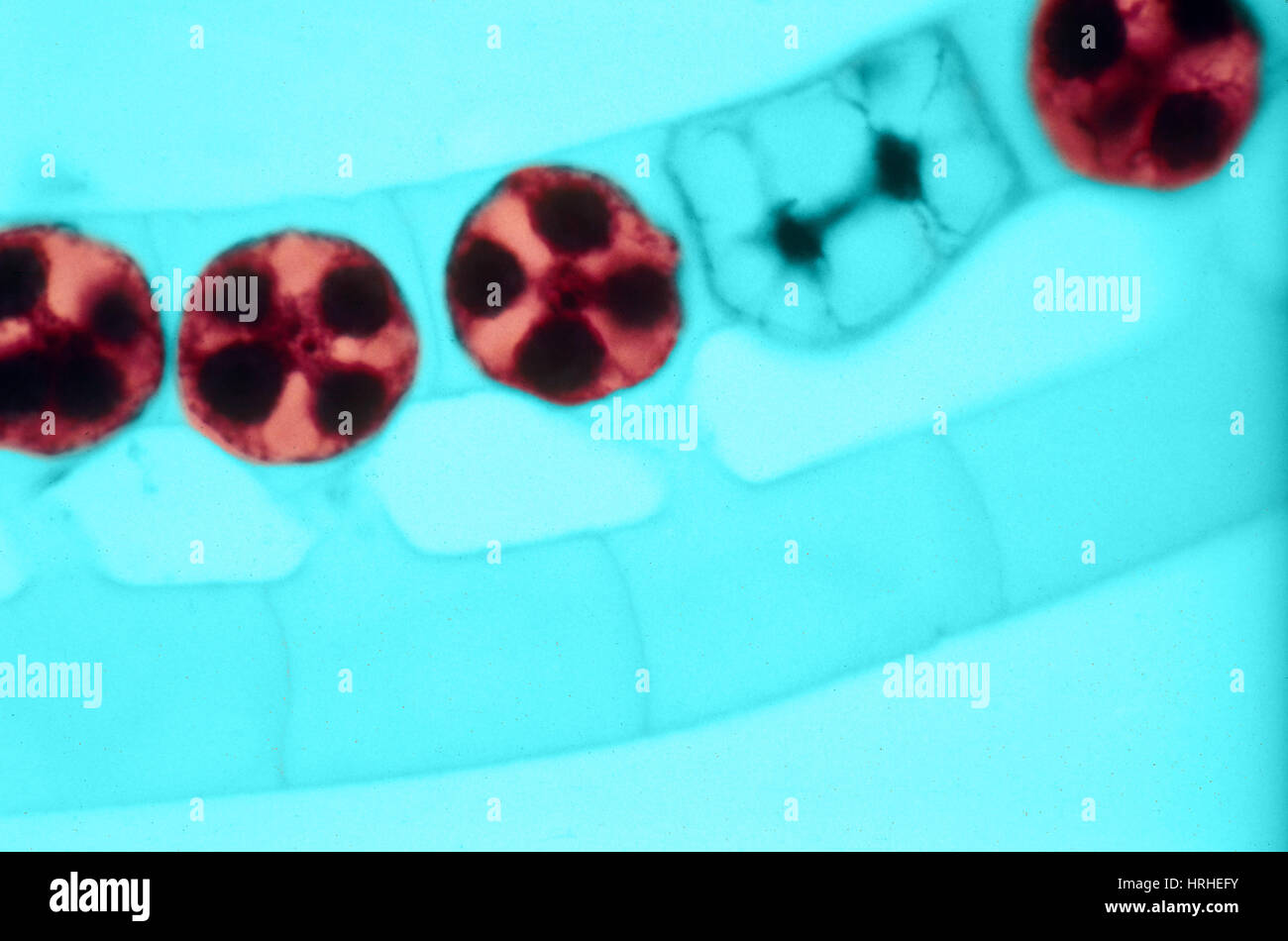
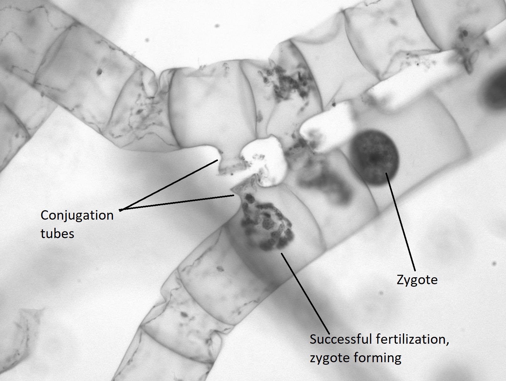
Spirogyra conjugating
Morphological Observation of Spirogyra ellipsospora Transeau, an Edible ... This study is the first report which focused on the morphology of S. ellipsospora, as an edible macroalgae. Background Spirogyra Link (1820) is an unbranched filamentous green alga that forms free ...

The effects of hydrogen-rich saline solution on intestinal ...
Genus Spirogyra - An Overview - Microbe Notes Spirogyra is a green alga that is mostly found in freshwater in the form of clumps. These are also called Water silk or Mermaid's tresses. It is a unicellular organism but can be seen in freshwater bodies as it clumps together to form a multicellular structure. Spirogyra consists of chlorophyll which gives it a green appearance.
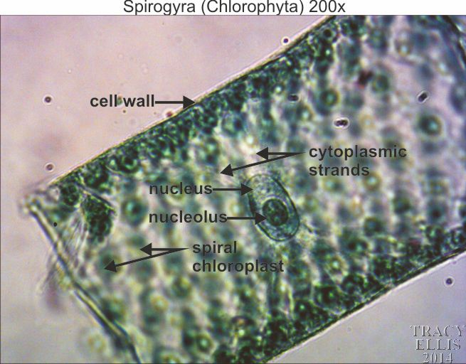
Spirogyra (chlorophyta) 200x - Dissection Connection
Compound Microscope- Definition, Labeled Diagram, Principle, Parts, Uses The optical microscope often referred to as the light microscope, is a type of microscope that uses visible light and a system of lenses to magnify images of small subjects. There are two basic types of optical microscopes: Simple microscopes. Compound microscopes. The term "compound" in compound microscopes refers to the microscope having ...
Plants | Free Full-Text | Induction of Conjugation and ...
Microscope Imaging Station. Gallery. - Exploratorium This algae grows in long, hairlike strands in freshwater ponds. The helical green structures inside the cell walls are chloroplasts. Filaments of the green algae Spirogyra were mounted in pondwater between a slide and coverslip using a silicon spacer. Images were taken on a compound inverted microscope using a 40x DIC objective and digital camera.

Spirogyra fresh water algae, microscope view Stock Photo - Alamy
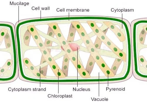
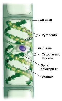
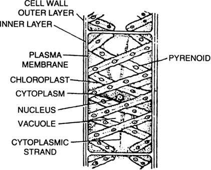
Draw a neat diagram of spirogyra and label on it: A cell of spirogyra comprises a cell wall, pyrenoids, nucleus, cytoplasmic threads, spiral chloroplasts, and vacuole. The labeled diagram of spirogyra is given below. In a microscopic view we can see the various parts of the cell of spirogyra. Cell wall is the outermost layer of a plant cell. Pyrenoid is a structure inside or beside the ...
The Genus Oedogonium
Diatoms Under A Microscope Labeled - chunyinga.blogspot.com Microscope labeled diagram 1. They are generally of a golden-brown color and many are able to move about. What Are Diatoms Diatoms Of North America. Elegans under a stereo microscope. Full Hd Live Diatom Algae Under Microscope Magnification 400x.
Lab Practical 101 Flashcards | Chegg.com
Spirogyra Flashcards | Quizlet Spirogyra Occurance mostly freshwater, floating pond silk common name for spirogyra due to its slippery touch structure multicellular, long filamentous body composed of many similar cells cell wall outermost covering made of cellulose with pectin on the outerside pectin gets sticky in water, causes spirogyra to be slippery cell membrane
4.6: Green Algae - Biology LibreTexts
The following diagram illustrates a filament of spirogyra as ... - YouTube The following diagram illustrates a filament of spirogyra as seen under the microscope. Its parts have been labelled as A,B,C,D. Functions of these parts are...

Spirogyra
Spirogyra Under Microscope 100X - Shutterstock Puzzlepix, Spirogyra ... Spirogyra Under Microscope 100X - Shutterstock Puzzlepix, ... Paramecium under microscope 400x labeled. Http Www Nyms Org Wp Content Uploads 2012 04 Newsletter 2016 03 Nyms Extended Email Rev1 Pdf from A d light microscopy images of nebela gimlii test a test. Not the most beautiful image ever, but it's representative of the diversity of shapes ...
Lateral Conjugation Spirogyra Algae Under Microscope Foto ...
Spirogyra: Structure, Diagram, Fragmentation, Sexual Reproduction - BYJUS Spirogyra are free-floating green algae present in freshwater habitats such as ponds, lakes, etc. Spirogyra are commonly known as "water silk or pond silk". They have a filamentous and unbranched vegetative structure. There are around 400 species of Spirogyra found.

Untitled Document
Labeled Diagram of Spirogyra - QS Study Spirogyra is a sophisticated, filamentous green alga, found in freshwater represented by about 300 species. It is also identified as pond silk, as its fiber burnishes like silk due to the occurrence of mucilage. The vegetative body structure of spirogyra. A) External features. The vegetative body of Spirogyra is unbranched and filamentous.

Microscopic photograph of Spirogyra sp. (40X) | Download ...
Spirogyra_diagram_labeled | Spirogyra diagram, Teaching biology, Middle ... Spirogyra_diagram_labeled Find this Pin and more on Algae by Aerobe. More like this Biology Projects Biology Art Biology Lessons Cell Biology School Projects Science Models Single Celled Wooden Main Door Design Green Algae C (914) 456-6009 Dna Microscopic Algae Microscopic Images Science Biology Flora Protists Bio Art Plant Cell

Microscope World Blog: Spirogyra under the Microscope
Chorophyta Spirogyra sp BY Dwi Kurnia Mei Lailatul
Filaments Spirogyra Alga Under Microscope Stock Photo ...
Spirogyra filament hi-res stock photography and images - Alamy

291 Algae Under Microscopic View Photos - Free & Royalty-Free ...

Lab: Cells and the Microscope - PDF Free Download
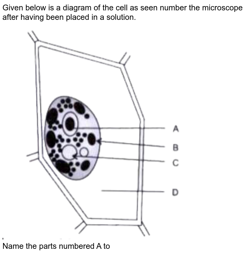
The following diagram illustrates a filament of spirogyra as ...

Keratella rotifer and Spirogyra algae, light micrograph ...

Well labelled Diagram of Spirogyra | What is Spirogyra | Simplest way of drawing the spirogyra

43787_Slide_64422_05222015_125 ...
Nature speaks about God: Algae and stars | Lynne Baab

Vegetative Spirogyra - Prepared Microscope Slide - 75x25mm ...

Microscopic View of Spirogyra sp. | Download Scientific Diagram
Spirogyra Sp Algae Under Microscopic View Foto Stok ...
4.6: Green Algae - Biology LibreTexts
Spirogyra Under a Light Microscope, 100x Magnification Stock ...
Mic-UK: Reflections on studying Spirogyra - a classic school ...

Spirogyra | microscopesandmonsters
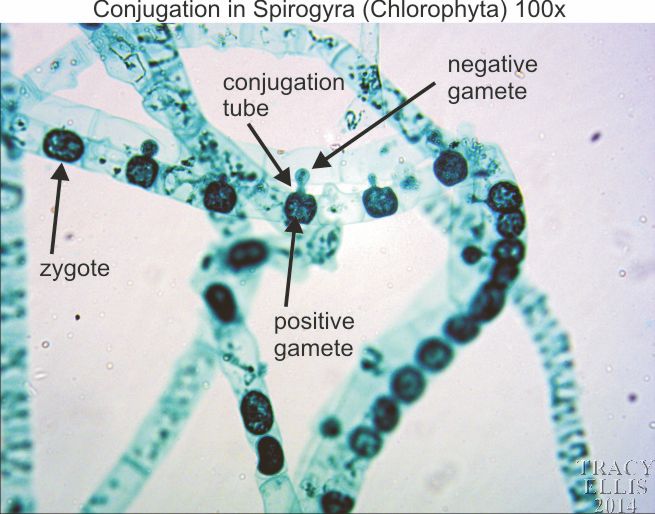
Spirogyra conjugation 100x - Dissection Connection
Labeled Diagram of Spirogyra - QS Study
Labeled Diagram of Spirogyra - QS Study

Microscopic View of Oscillatoria sp. | Download Scientific ...

Spirogyra - Wikipedia

Structure and Life Cycle of Spirogyra

Spirogyra Stock Illustrations – 52 Spirogyra Stock ...

File:Spirogyra Under Light Microscope.jpg - Wikimedia Commons

Spirogyra_diagram_labeled | Spirogyra diagram, Teaching ...
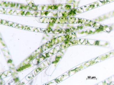
Figure 1 | Culture of Spirogyra sp. in a flat-panel airlift ...
Draw a neat diagram of spirogyra and label on it Dark class 9 ...

File:Spirogyra Under Light Microscope 40 X Magnification.jpg ...
Spirogyra: Structure & Characteristics with Labeled Diagram
Draw a labelled diagram of spirogyra cell. from Science ...

Superior carbon belts from Spirogyra for efficient ...
Post a Comment for "45 spirogyra microscope labeled"