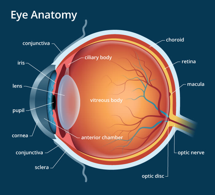
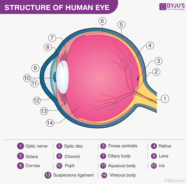
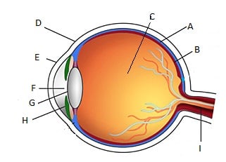
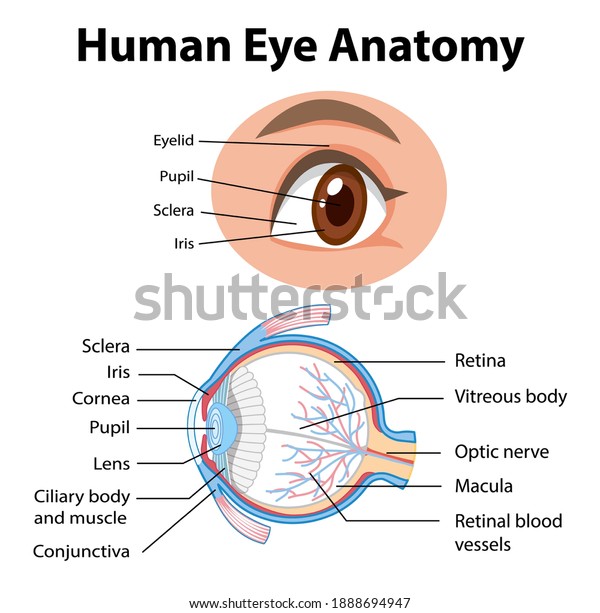
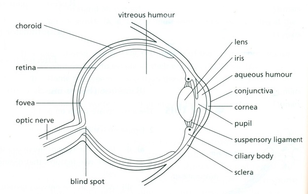
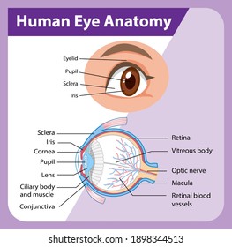
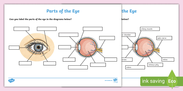
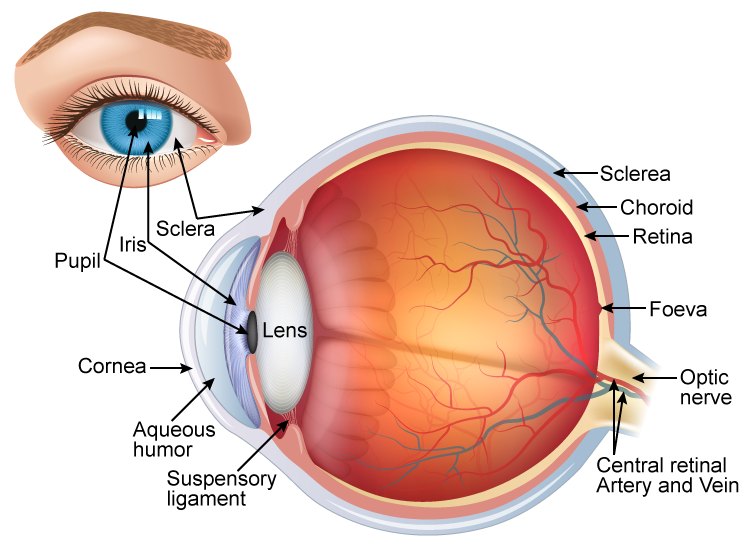
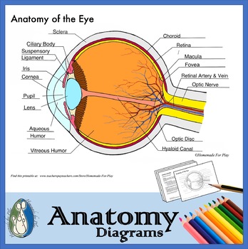
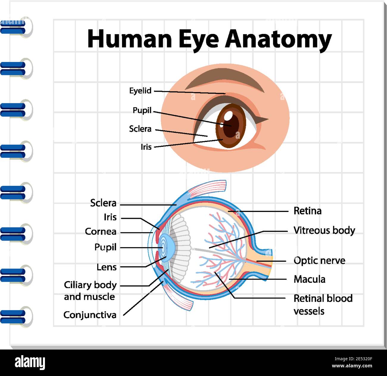
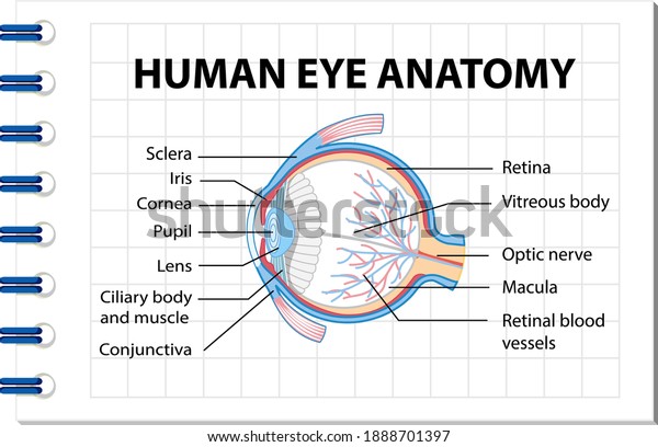
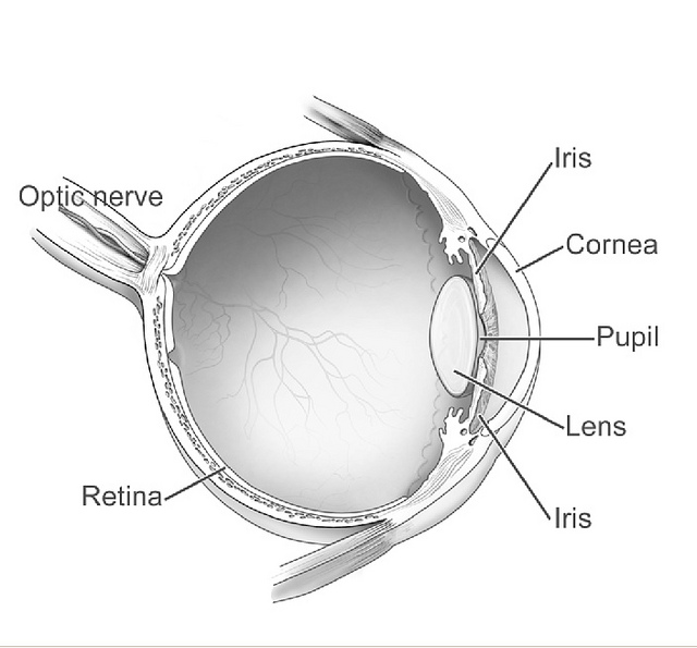
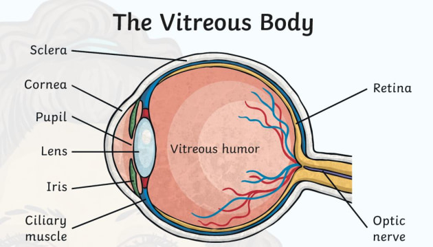
38 diagram of an eye with labels
Jupiter - Wikipedia Jupiter is the fifth planet from the Sun and the largest in the Solar System.It is a gas giant with a mass more than two and a half times that of all the other planets in the Solar System combined, but slightly less than one-thousandth the mass of the Sun. Jupiter is the third brightest natural object in the Earth's night sky after the Moon and Venus, and it has been observed since prehistoric ... Toolbox Talk Templates: Free Download | SafetyCulture Toolbox Talk Template. A toolbox talk template is used to document daily safety discussions prior to the work shift. Use this toolbox talk form to document a summary of the toolbox topic discussed and gather electronic signatures from workers present in the meeting. Maximize the use of this checklist by following the points below.
Azure icons - Azure Architecture Center | Microsoft Docs In diagrams, we recommend to include the product name somewhere close to the icon. Use the icons as they would appear within Azure. Don'ts. Don't crop, flip or rotate icons. Don't distort or change icon shape in any way. Don't use Microsoft product icons to represent your product or service. Example architecture diagram
:background_color(FFFFFF):format(jpeg)/images/library/11201/overview_parts_of_the_eye_labelled_diagram.jpg)
Diagram of an eye with labels
Without Egg, Sperm or Womb: Synthetic Embryo Models May Enable Growing ... A diagram showing the innovative method for growing synthetic mouse embryo models from stem cells - without egg, sperm or womb - developed in the laboratory of Prof. Jacob Hanna. Credit: Weizmann Institute of Science. Reference: "Post-Gastrulation Synthetic Embryos Generated Ex Utero from Mouse Naïve ESCs" by Shadi Tarazi, Alejandro ... Eye anatomy: Muscles, arteries, nerves and lacrimal gland - Kenhub Bony cavity within the skull that houses the eye and its associated structures (muscles of the eye, eyelid, periorbital fat, lacrimal apparatus) Bones of the orbit. Maxilla, zygomatic bone, frontal bone, ethmoid bone, lacrimal bone, sphenoid bone and palatine bone. Structure of the eye. Cornea, anterior chamber, lens, vitreous chamber and ... Nerve Supply Of The Jaws And Teeth - Dental Anatomy (1) The maxillary teeth. The maxillary nerve on each side passes forward in the floor of the orbit of the eye. It first gives off the posterior superior alveolar branch to the three maxillary molars. When in the floor, the maxillary nerve gives off a middle superior alveolar branch to the maxillary bicuspids and mesial root of the first molar.
Diagram of an eye with labels. Human Ear Diagram Without Labels - picture front of the eye without ... Human Ear Diagram Without Labels - 18 images - datei anatomy of the human ear svg wikipedia, ear anatomy diagram, circulatory system diagram without labels fresh smorgasbord variety, unlabeled digestive system diagram without labels news word, Angiography: Purpose and How It's Done - Verywell Health The angiography procedure involves several steps: After creating a small incision, a sheath is inserted into the blood vessel which allows for the insertion of the guidewire and catheter, as well as the injection of contrast medications. The guidewire is visible with X-ray and can be tracked as it progresses through the circulatory system. Common Assistive Technologies - Blind/Visual Impairment - LibGuides at ... The Technology Related Assistance to Individuals with Disabilities Act of 1988 described an assistive technology device as "any item, piece of equipment, or product system, whether acquired commercially off the shelf, modified, or customized, that is used to increase, maintain, or improve functional capabilities of individuals with disabilities." ... SQL exercises on employee Database - w3resource SQL employee Database [115 Exercise with Solution] [An editor is available at the bottom of the page to write and execute the scripts.Structure of employee Database: 1. From the following table return complete information about the employees.
Parts of Human Eye and Their Functions | MD-Health.com The iris is the area of the eye that contains the pigment which gives the eye its color. This area surrounds the pupil, and uses the dilator pupillae muscles to widen or close the pupil. This allows the eye to take in more or less light depending on how bright it is around you. If it is too bright, the iris will shrink the pupil so that they ... WHMIS 2015 - Labels : OSH Answers Suppliers and employers must use and follow the WHMIS 2015 requirements for labels and safety data sheets (SDSs) for hazardous products sold, distributed, or imported into Canada. Please refer to the following other OSH Answers documents for more information: WHMIS 2015 - General. WHMIS 2015 - Pictograms. Cell biology Virtual Lab I - Amrita Vishwa Vidyapeetham The objective lens at first formed a real and inverted magnified image. And then the eye piece further magnifies the same image to virtual magnified image. Focusing On Microscopic Objects . Start with Clean Lenses: It is important that microscope lenses be very clean. Before viewing through a microscope, use lens paper to gently clean the lenses. Structure and parts of a sperm cell - invitra.com This labelled diagram shows the structure of a sperm cell in detail, which has the following parts: Head With its spheric shape, it consists of a large nucleus, which at the same time contains an acrosome. The nucleus contains the genetic information and 23 chromosomes. It also secretes a hyaluronidase enzyme that destroys the hyaluronic acid ...
Histone - Genome.gov A histone is a protein that provides structural support for a chromosome. Each chromosome contains a long molecule of DNA, which must fit into the cell nucleus. To do that, the DNA wraps around complexes of histone proteins, giving the chromosome a more compact shape. Histones also play a role in the regulation of gene expression. Foot Anatomy and Common Foot Problems - Verywell Health Plus, the foot must be flexible to adapt to uneven surfaces and remain stable. Common foot problems include plantar fasciitis, bunions, flat feet, heel spurs, mallet toe, metatarsalgia, claw toe, and Morton's neuroma. This article provides an overview of foot anatomy and foot problems that come from overuse, injury, and normal wear and tear of ... What Are the Most Sensitive Areas in Women? - New Health Advisor 3. Nipples. Nipples are one of the most well-known places among the 5 most sensitive parts on the female body. There are ways to utilize them to their maximum potential. How to do: Cupping a breast in your hand while gently touching or licking regions around the nipples will slowly stimulate the body. Then turn your focus to the nipples by ... Phonograph record - Wikipedia A phonograph record (also known as a gramophone record, especially in British English), or simply a record, is an analog sound storage medium in the form of a flat disc with an inscribed, modulated spiral groove. The groove usually starts near the periphery and ends near the center of the disc. At first, the discs were commonly made from shellac, with earlier records having a fine abrasive ...
Anatomy of the Ear - Audiology Net Nice site that shows drawn pictures of the cranial nerves. It also explains some topics like Bell's Palsy. Anatomy of the Ear and Animations. Click on animations and see how the ear works. Auditory and Vestibular Pathways. Great site with a good combination of pictures and information. Temporal Bone Anatomy web site.
Research Guides: APA Citation Style, 7th edition: Figures Figures should be labeled "Figure (number)" ABOVE the figure. Double-space the caption that appears under a figure. General Format 1 (Figure from a Book): ... 2300 Eye Street, NW Washington, DC 20037. 202-994-2850. Website. Social: Facebook Page Twitter Page YouTube Page << Previous: Electronic Image;
be quiet! Pure Base 500 FX Mid-Tower Smart Chassis Review The front I/O panel offers USB 3.2 Gen. 2 Type-C and USB 3.2 Gen. 1 ports for connectivity to other devices. We also get a power button and an ARGB switch, along with a pair of 3.5mm HD Audio ...
Brain, spinal cord and peripheral nervous system anatomy - Kenhub Diagram of the brain (labeled) The most superficial layer of the cerebrum is the cerebral cortex.It is a layer of grey matter which displays numerous folds (sulci and gyri), can be categorization structurally (cortical cytoarchitecture) or functionally (Brodmann areas), and is home to areas such as the primary motor cortex and the primary somatosensory cortex, both of which house a homunculus.
Spectrophotometry (Theory) - Amrita Vishwa Vidyapeetham Diagram of Beer-Lambert absorption of a beam of light as it travels through a cuvette of width . Beer-Lambert's law is the linear relationship between the absorbance and concentration of the absorbing sample, i.e. a logarithmic relation exist between the transmission of light through a substance () ...
WHMIS 2015 - Pictograms : OSH Answers Pictograms are graphic images that immediately show the user of a hazardous product what type of hazard is present. With a quick glance, you can see, for example, that the product is flammable, or if it might be a health hazard. Most pictograms have a distinctive red "square set on one of its points" border.
Circulatory System Diagram - New Health Advisor This circuit typically includes the movement of blood inside heart and 'myocardium' (the membrane of heart). Coronary circuit mainly consists of cardiac veins including anterior cardiac vein, small vein, middle vein and great (large) cardiac vein. There are different types of circulatory system diagrams; some have labels while others don't.
APA Citation Style, 7th edition - George Washington University drawings. photographs/images. This section will cover the following examples: Image from an Electronic Source. Figures. For more examples and information, consult the following publications: Publication Manual of the American Psychological Association (7th ed.) Call Number: BF76.7 .P83 2020.
Positions and Functions of the Four Brain Lobes - MD-Health.com The occipital lobe, the smallest of the four lobes of the brain, is located near the posterior region of the cerebral cortex, near the back of the skull. The occipital lobe is the primary visual processing center of the brain. Here are some other functions of the occipital lobe: Visual-spatial processing. Movement and color recognition.
Explore: Almost 14,000 eviction notices served in SF public housing ... Explore: Thousands of eviction notices served in SF public housing over 5 years. Property managers say they struggle to provide for tenants with increasingly complex needs, leading to thousands of eviction proceedings. by Will Jarrett August 2, 2022. The Henry Hotel, operated by Caritas Management, saw 37 evictions between 2016 and 2020 - the ...
Meiosis - Genome.gov Meiosis is a type of cell division in sexually reproducing organisms that reduces the number of chromosomes in gametes (the sex cells, or egg and sperm). In humans, body (or somatic) cells are diploid, containing two sets of chromosomes (one from each parent). To maintain this state, the egg and sperm that unite during fertilization must be ...
Nerve Supply Of The Jaws And Teeth - Dental Anatomy (1) The maxillary teeth. The maxillary nerve on each side passes forward in the floor of the orbit of the eye. It first gives off the posterior superior alveolar branch to the three maxillary molars. When in the floor, the maxillary nerve gives off a middle superior alveolar branch to the maxillary bicuspids and mesial root of the first molar.
Eye anatomy: Muscles, arteries, nerves and lacrimal gland - Kenhub Bony cavity within the skull that houses the eye and its associated structures (muscles of the eye, eyelid, periorbital fat, lacrimal apparatus) Bones of the orbit. Maxilla, zygomatic bone, frontal bone, ethmoid bone, lacrimal bone, sphenoid bone and palatine bone. Structure of the eye. Cornea, anterior chamber, lens, vitreous chamber and ...
Without Egg, Sperm or Womb: Synthetic Embryo Models May Enable Growing ... A diagram showing the innovative method for growing synthetic mouse embryo models from stem cells - without egg, sperm or womb - developed in the laboratory of Prof. Jacob Hanna. Credit: Weizmann Institute of Science. Reference: "Post-Gastrulation Synthetic Embryos Generated Ex Utero from Mouse Naïve ESCs" by Shadi Tarazi, Alejandro ...













Post a Comment for "38 diagram of an eye with labels"